
Master’s Degree Awarded to Ms. Dania Naji Al-Sari in Oral and Maxillofacial Surgery
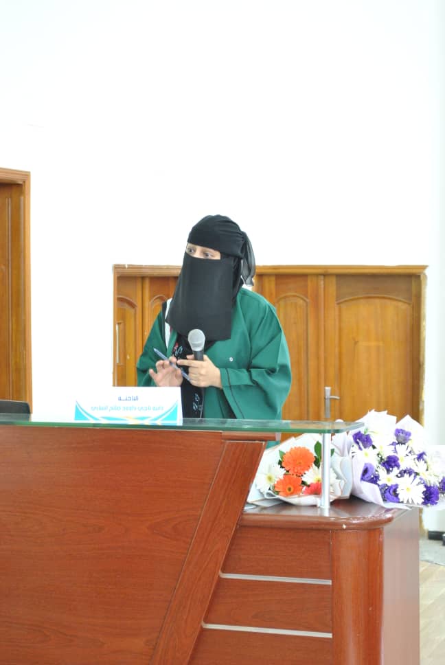
Ms. Dania Naji Dawood Saleh Al-Sari was awarded a Master’s Degree in Oral and Maxillofacial Surgery for her thesis titled: Cone-Beam Computed Tomography Evaluation of Lingual Foramen and Canal among a Sample of Yemeni Population, which was submitted to the Department of Oral and Maxillofacial Surgery, Faculty of Dentistry– Sana’a University. The MA defense was held on Wednesday, April 22, 2026 .
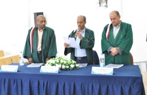
The MA Viva-voce Committee, which was formed based on a resolution issued by the Graduate Studies and Scientific Research Council, consisted of the following:
| # | Committee Members | Designation | Position |
| 1 | Prof. Sam Abdulkarim Da’er | Internal Examiner | Chair |
| 2 | Prof. Akram Thabet Nasher | Main Supervisor | Member |
| 3 | Asst. Prof. Nashwan Hameed Saleh Al-Tairi | External Examiner | Member |
The thesis aimed to:
- Evaluate the mandibular lingual foramina and their canals, and investigate their anatomical variations in a sample of Yemeni adults using cone-beam computed tomography (CBCT).
The study adopted a retrospective cross-sectional descriptive design conducted on 200 CBCT scans (90 males and 110 females) with a mean age of 34.6 ± 11.2 years, obtained from radiology centers in Sana’a. CBCT images were acquired using the EZ3D-I system (Vatech, Korea) with a slice thickness of 0.5 mm. The presence of median lingual foramina (MLF) and lateral lingual foramina (LLF), along with their canals, number, location, diameter, and linear measurements, were assessed. Associations with gender and age groups (<30, 30–39, 40–49, ≥50 years) were analyzed. Data were analyzed using SPSS version 26, employing Kruskal–Wallis, Mann–Whitney, Chi-square, and Welch tests, with statistical significance set at p < 0.05.
The study yielded several key findings summarized as follows:
- All CBCT scans (100%) showed at least one lingual foramen with an associated canal.
- A total of 885 foramina and canals were identified, with 44.5% being median and 55.5% lateral.
- Detection rates were 99% for median foramina and 94% for lateral foramina, with higher prevalence among females.
- Statistically significant gender differences were observed in the maximum number of lateral foramina, with two being most common in males and three in females.
- Most lateral foramina (76.5%) were located in the incisor–canine region, with higher prevalence among females.
- In 52% of cases, foramina diameter exceeded 1 mm, more frequently in males. Median foramina were generally >1 mm, whereas lateral foramina were typically ≤1 mm.
- Significant differences were observed across gender and age groups in several linear measurements, with clear distinctions between median and lateral foramina.
- The study concluded that Median and lateral lingual foramina of the mandible are common anatomical structures, with prevalence rates of 99% and 94%, respectively.
In light of these findings, the researcher recommended the following:
- Careful evaluation by dentists and oral and maxillofacial surgeons during surgical procedures.
- Routine preoperative assessment using CBCT to accurately identify the location and dimensions of lingual foramina and canals in order to avoid potential neurovascular complications.
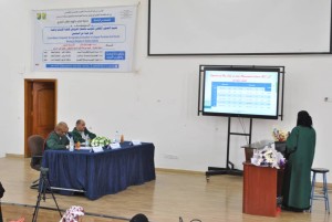
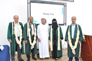
The defense session was attended by a number of academics, researchers, students, colleagues, and the researcher’s family.

")

")